Oval Shapped Material Inside of a Fecal Reading in Dogs
Epithelial and white blood cells in stool.
Epithelial and white blood cells are often seen in trichrome-stained stool smears and may be mistaken for amebae.
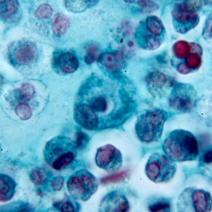
Effigy A: White blood cells in a trichrome-stained stool smear.
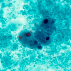
Effigy B: Macrophages in a trichrome-stained stool smear.
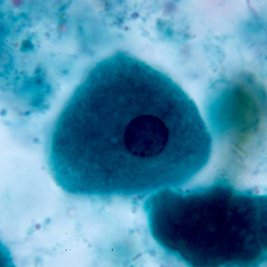
Figure C: Epithelial cell in a trichrome-stained stool smear.
Yeast and other fungal elements in stool.
Yeast and other fungal elements are mutual in stool. Depending on the size and shape, they may be confused for a variety of helminth and protozoan species.
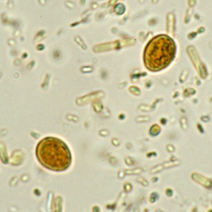
Figure A: Yeast in an iodine-stained full-bodied wet mountain of stool. Yeast in wet mounts may exist confused for Giardia.

Effigy E: Fungal spore in a wet mountain of stool. Such spores may be dislocated for the cysts of Entamoeba spp.
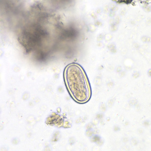
Figure B: Spore of a morel mushroom. Such spores may be dislocated for helminth eggs, specially hookworm.

Effigy C: Fungal spore in concentrated wet mounts of stool. Such spores may exist confused for protozoa such as Giardia or Chilomastix.
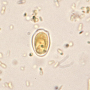
Figure D: Fungal spore in concentrated wet mounts of stool. Such spores may be dislocated for protozoa such equally Giardia or Chilomastix.
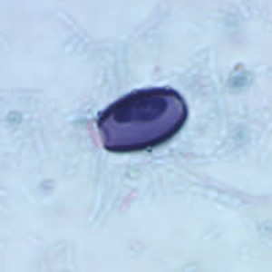
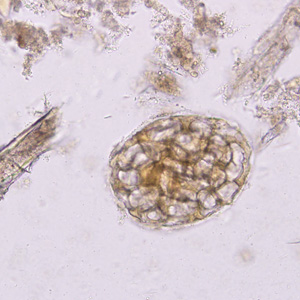
Pollen grains in stool.
Pollen spores are very common in stool and may be dislocated for a diversity of helminth eggs.

Figure A: Pollen grain in a concentrated wet mount of stool. This grain looks very similar to the fertile egg of Ascaris lumbricoides, although the spine-like structures on the outer layer should carve up the two.
Effigy Due east: Possible pollen grain or algal or fungal spore, similar to the 1 in Figure B, but in a trichrome-stained stool specimen. Image courtesy of the Washington Country Public Health Laboratories.

Effigy B: Possible pollen grain or algal or fungal spore in a full-bodied wet mountain of stool. Grains like this 1 resemble the operculated eggs of Clonorchis, Metagonimus and related. These grains are commonly smaller than the trematode eggs, nevertheless. Paradigm courtesy of the Washington State Public Health Laboratories.
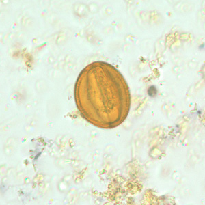
Figure C: Pollen grain in a concentrated wet mount of stool.
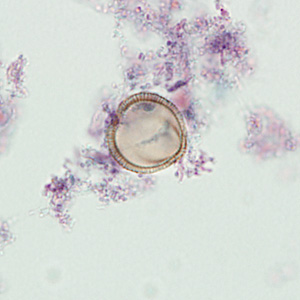
Figure D: Pollen grain in a trichrome-stained stool specimen. In this focal plane, the grain looks like the striated egg of Taenia sp. However, observe the lack of refractile hooks.
Constitute material and constitute hairs in stool.
Institute material is very common in stool and may be dislocated for a variety of helminth eggs and larvae.
Effigy A: Plant cell in a concentrated wet mountain of stool. Such fabric can be common in stool and may be confused for helminth eggs, although they are commonly much larger than the eggs of nearly helminth species.
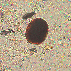
Figure B: Plant textile in an iodine-stained full-bodied wet mount of stool. This material can exist confused for a hookworm egg. Image courtesy of the Alaska State Public Health Laboratory.

Effigy C: Plant hair in a concentrated wet mount of stool. Plant hairs can exist common in stool and may exist confused for the larvae of hookworm or Strongyloides stercoralis. However, they are often cleaved at one end, take a refractile center and lack the strictures seem in helminth larvae (esophagus, genital primordium, etc).
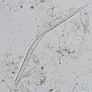
Figure D: Establish pilus in a concentrated wet mount of stool. Establish hairs can be common in stool and may exist confused for the larvae of hookworm or Strongyloides stercoralis. Yet, they are often broken at ane cease, accept a refractile middle and lack the strictures seem in helminth larvae (esophagus, genital primordium, etc).
Artifacts in acid-fast stained stool specimens.
Several objects may be seen in acrid-fast stained stool specimens that may be confused for the oocysts of Cryptosporidium and Cyclospora.
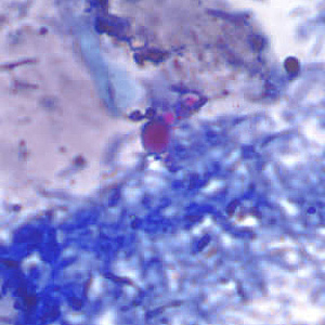
Effigy A: Yeast in an acid-fast stained stool specimen. These may be confused for the oocysts of Cryptosporidium sp. Images courtesy of the Arizona State Public Wellness Laboratory.

Figure East: Object, probably fungal, in an acid-fast stained stool specimen. Such objects may be confused for the oocysts of Cyclospora spp.
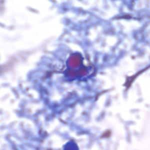
Figure B: Yeast in an acrid-fast stained stool specimen. These may exist dislocated for the oocysts of Cryptosporidium sp. Images courtesy of the Arizona State Public Wellness Laboratory.

Effigy F: Object, probably fungal, in an acid-fast stained stool specimen. Such objects may exist dislocated for the oocysts of Cyclospora spp.
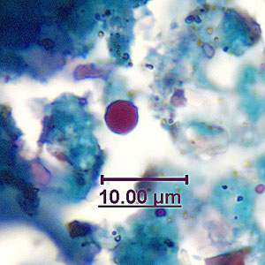
Figure C: Fungal element in an acid-fast stained stool specimen. Such objects may exist confused for the oocysts of Cryptosporidium spp. Images courtesy of the Georgia State Public Wellness Laboratory.
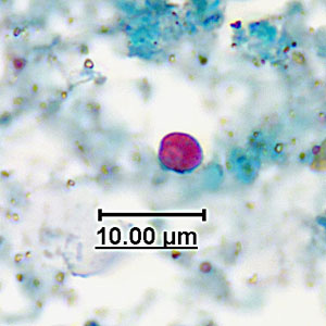
Figure D: Fungal element in an acid-fast stained stool specimen. Such objects may exist confused for the oocysts of Cryptosporidium spp. Images courtesy of the Georgia State Public Health Laboratory.
Miscellaneous artifacts in stool.
Miscellaneous artifacts in stool.

Figure A: Unknown object in a concentrated stool specimen. This object looks like the egg of Hymenolepis but lacks refractile hooks and the polar filaments seen in H. nana.
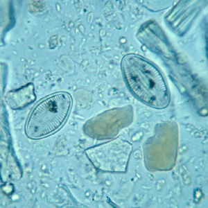
Figure East: Diatoms in stool. Diatoms do not specifically resemble whatsoever parasites of humans, only their size and shape and apparent structure may cause the microscopist to take notice.
Effigy B: Unknown objects in a formalin-concentrated stool specimen. Although these objects do not specifically resemble whatsoever parasites of humans, their unique appearance may cause the microscopist to take find.
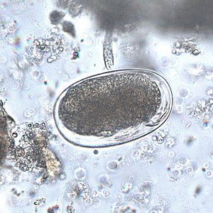
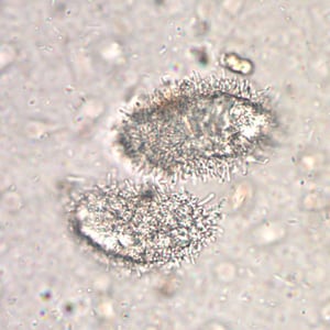
Effigy F: Mite egg in a formalin-concentrated stool specimen. Mite eggs are similar to hookworm eggs simply are usually larger (just not always). In this specimen, leg buds can exist seen in the lower right area of the egg.
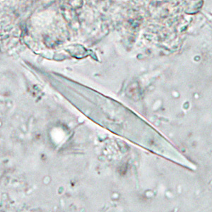
Figure C: Charcot-Leyden crystals. These crystals are the breakdown products of eosinophils and maybe found in the feces and sputum of people with tissue-invading parasitic infections or diverse allergic reactions.
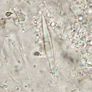
Figure D: Charcot-Leyden crystals. These crystals are the breakdown products of eosinophils and perhaps constitute in the carrion and sputum of people with tissue-invading parasitic infections or various allergic reactions.
Platelets.
Elongated and degenerating platelets in blood may be confused for Trypanosoma spp. or malaria elements.

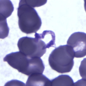
Figure A: Platelet in a thick claret smear. The nature of the platelet gives it the appearance of a trypomastigote of Trypanosoma sp.

Figure B: Platelet in a sparse claret smear. The nature of the platelet gives it the appearance of a trypomastigote of Trypanosoma sp.

Effigy C: Platelets in thin claret smears. The nature of the platelets gives them the appearance of trypomastigotes of Trypanosoma cruzi.
Effigy D: Platelets in sparse blood smears. The nature of the platelets gives them the appearance of trypomastigotes of Trypanosoma cruzi.
White blood cells.
Normal white blood cells.
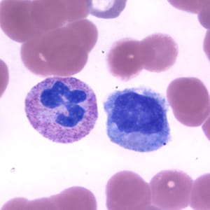
Figure A: Neutrophil (left) and monocyte (right) in a thin blood smear, stained with Giemsa.
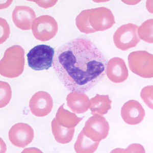
Figure East: Small lymphocyte (left) and neutrophil in a thin blood smear, stained with Giemsa.
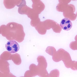
Effigy B: Neutrophils in a sparse blood smear, stained with Giemsa.
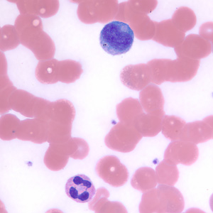
Figure F: Neutrophil (lower) and lymphocyte (upper) in a thin blood smear, stained with Giemsa.
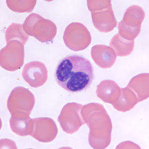
Figure C: Neutrophil in a thin blood smear, stained with Giemsa.
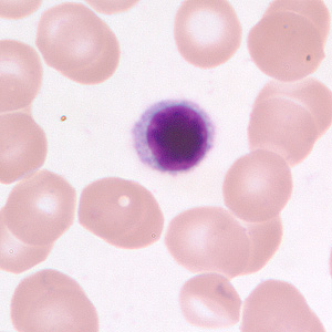
Figure D: Small lymphocyte in a thin blood smear, stained with Giemsa.
Miscellaneous blood elements.
Miscellaneous blood elements.

Figure A: Unidentified objects (probably fungal) in a thin blood smear, stained with Giemsa. Such objects may be confused for amastigotes of Leishmania or Trypanosoma cruzi, but the lack of a singled-out nucleus and kinetoplast should dominion-out these parasites.
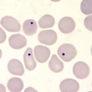
Figure East: Howell-Jolly bodies in a thin claret smear, stained with Giemsa. Howell-Jolly bodies are inclusion that may be seen in splenectomized patients or patients with an otherwise non-functioning or atrophic spleen, and in patients with astringent anemia or leukemia.
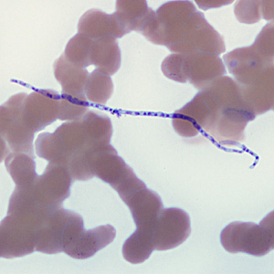
Figure B: Fungal spore of Helicosporium (or related). Such objects are air-borne contaminants in laboratories and may be mistaken for microfilariae in stained blood smears.
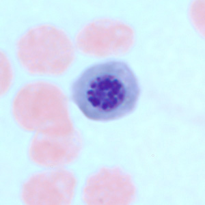
Figure C: Nucleated ruddy claret cell in a thin claret smear, stained with Giemsa. There are several atmospheric condition which tin cause a premature release of nucleated red blood cells into apportionment. Such objects may exist confused for schizonts of Plasmodium spp.
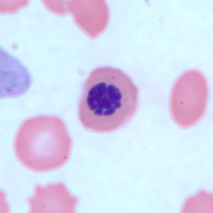
Figure D: Nucleated ruby blood cell in a thin blood smear, stained with Giemsa. In that location are several conditions which tin cause a premature release of nucleated red blood cells into apportionment. Such objects may be dislocated for schizonts of Plasmodium spp.
Miscellaneous artifacts in tissue.
Miscellaneous blood and tissue elements.

Effigy A: Yeast in a Giemsa-stained tissue biopsy. Such objects may be confused for amastigotes of Leishmania or Trypanosoma cruzi, but the lack of a distinct nucleus and kinetoplast should rule-out these parasites.

Figure B: Seed in an abdominal biopsy specimen. Such object may be confused with parasites, such as intestinal trematodes or Balantidium coli. The boxy, compartmentalized cells are characteristic of institute tissue. Image courtesy of the Children's Hospital of Eastern Ontario, Canada.
Earthworms, horsehair worms and aquatic wing larvae.
Occasionally, not-parasitic worms and insects are brought to the laboratory for identification. They need to be distinguished from truthful parasites and vectors.

Figure A: Earthworms (Lumbricus and related) are normally sent to the public health laboratories for identification. The presence of setae, segmentation, and a clitellum (red pointer, Figure B) should distinguish them from parasitic helminths. Images courtesy of the Kentucky Department of Health.
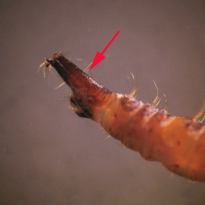
Figure E: Aquatic larvae of flies. The free-living aquatic larvae of diverse flies breed in standing water, including toilets, leading to the misconception they came from stool or urine. The presence of prolegs, a caput capsule, breathing tubes (arrow), division and/or setae will usually distinguish them from true parasitic worms.
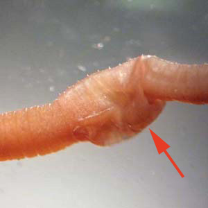
Figure B: Earthworms (Lumbricus and related) are ordinarily sent to the public health laboratories for identification. The presence of setae, division, and a clitellum (red arrow) should distinguish them from parasitic helminths. Images courtesy of the Kentucky Section of Health.
Effigy F: Aquatic larvae of flies. The free-living aquatic larvae of various flies breed in continuing water, including toilets, leading to the misconception they came from stool or urine. The presence of prolegs, a head capsule, animate tubes (arrow, Figure E), segmentation and/or setae will normally distinguish them from truthful parasitic worms.

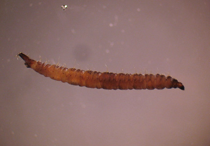
Figure C: Horsehair worms (Gordius and related). Horsehair worms are parasites of insects and may be found in households and end up in toilets. Every bit a outcome, they are ofttimes sent to the public wellness laboratories for identification. Images courtesy of the Wisconsin State Laboratory of Hygiene.
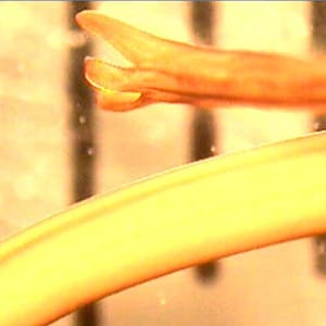
Figure D: Horsehair worms (Gordius and related). Horsehair worms are parasites of insects and may be found in households and finish up in toilets. Equally a result, they are oftentimes sent to the public health laboratories for identification. Images courtesy of the Wisconsin State Laboratory of Hygiene.
Source: https://www.cdc.gov/dpdx/artifacts/index.html
0 Response to "Oval Shapped Material Inside of a Fecal Reading in Dogs"
Post a Comment